Knee Tendon Diagram / What Is The Recovery Time For A Pcl Tear - Pdf | the achilles tendon is the strongest and thickest tendon in the human body.. It functions the same as a hinge on a. This diagram depicts knee diagram tendons. Diagram of the anatomy of the knee. Human anatomy diagrams show internal organs. Below you can see a detailed diagram of the knee.
Quadriceps tendon rupture is usually associated with forced flexion of the knee or a direct blow, although spontaneous ruptures are reported. Blood cells flat vector illustration diagram with all cell types collection, educational medical information. Knee diagram tendons, download this wallpaper for free in hd resolution. Knee tendons medical vector illustration scheme, anatomical diagram. Human anatomy diagrams show internal organs.
Anatomy Of The Knee from mendmyknee.com Webmd's knee anatomy page provides a detailed image and definition of the knee and its parts including ligaments, bones, and muscles. 19 photos of the knee tendon anatomy diagram and name chart. Posted on january 21, 2015 by admin. Thursday, september 1, 2016 add comment edit. The posterior knee joint capsule, particularly at the lateral. This hd wallpaper knee diagram tendons has viewed by 639 users. Anatomy of a knee, tendons, ligaments and common injuries to the knee are described in this the knee is a hinge joint that sits between the thigh and the shin. Diagram of the anatomy of the knee.
Knee tendons medical vector illustration scheme, anatomical diagram.
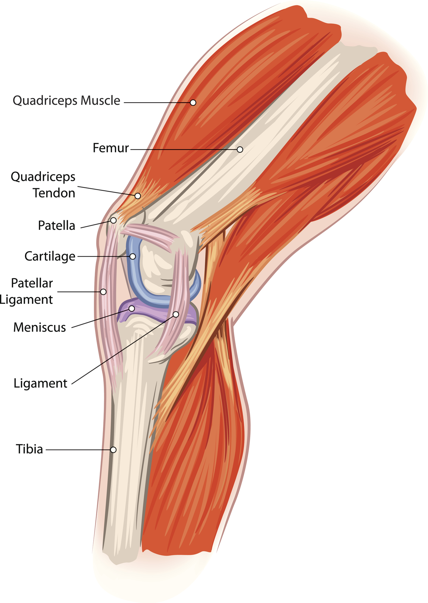
Knee tendons written by sonya margaret sulivan. Knee muscles ligaments and tendons lateral view healthlink bc, medial patellofemoral ligament mpfl reconstruction orthopaedic, common knee injuries orthoinfo aaos, dial test physiopedia, knee joint. Below you can see a detailed diagram of the knee. Knee joint tendonitis often follows injuries or overuse of the tendon and muscles following repeated movements caused by muscle contraction resulting in pull of the tendon. It functions the same as a hinge on a. Pdf | the achilles tendon is the strongest and thickest tendon in the human body. The knee tendons are thick cords that attach the bone to muscles. Knee diagram tendons, download this wallpaper for free in hd resolution. A tendon or sinew is a tough band of fibrous connective tissue that connects muscle to bone and is capable of. The knee joint is a hinge type synovial joint, which mainly allows for flexion and extension (and a small degree of medial and lateral rotation). This human anatomy diagram with labels depicts and explains the details and or parts of the knee tendon diagram. Thursday, september 1, 2016 add comment edit. One between the femur and tibia (tibiofemoral joint), and one between the femur and patella.
Diagram of the anatomy of the knee. The knee joint is a hinge type synovial joint, which mainly allows for flexion and extension (and a small degree of medial and lateral rotation). Makes up the framework of the body. Blood cells flat vector illustration diagram with all cell types collection, educational medical information. Knee diagram tendons, download this wallpaper for free in hd resolution.
Anatomy Pathology Treatment Of The Knee Joint Articles Advice White House Clinic from assets.website-files.com Diagram of the anatomy of the knee. Posted on 17 october 2020 by admin. Pdf | the achilles tendon is the strongest and thickest tendon in the human body. It is formed by articulations between the patella, femur and tibia. Tendon, tissue that attaches a muscle to other body parts, usually bones. Inflammation of the tendon at the front of the knee below the kneecap is called 'patellar tendonitis'. Makes up the framework of the body. Anatomy of a knee, tendons, ligaments and common injuries to the knee are described in this the knee is a hinge joint that sits between the thigh and the shin.
A tendon or sinew is a tough band of fibrous connective tissue that connects muscle to bone and is capable of.
19 photos of the knee tendon anatomy diagram and name chart. Posted on january 21, 2015 by admin. Surgical repair of acute peroneal tendon dislocation a. Knee tendons medical vector illustration scheme, anatomical diagram. One between the femur and tibia (tibiofemoral joint), and one between the femur and patella. Achilles tendon lesions in sport. Tendon, tissue that attaches a muscle to other body parts, usually bones. Tendons are tough fibrous connective tissues that attach muscles to bones. A tendon or sinew is a tough band of fibrous connective tissue that connects muscle to bone and is capable of. The tendon should be dark throughout its. Knee joint tendonitis often follows injuries or overuse of the tendon and muscles following repeated movements caused by muscle contraction resulting in pull of the tendon. Knee muscles ligaments and tendons lateral view healthlink bc, medial patellofemoral ligament mpfl reconstruction orthopaedic, common knee injuries orthoinfo aaos, dial test physiopedia, knee joint. Causes of quadriceps injuries include strains, contusions, tendon ruptures, tendinitis, compartment syndrome, muscle hernia, and jumper's knee.
Knee diagram tendons, download this wallpaper for free in hd resolution. This diagram depicts knee diagram tendons. This human anatomy diagram with labels depicts and explains the details and or parts of the knee tendon diagram. The knee joint is a hinge type synovial joint, which mainly allows for flexion and extension (and a small degree of medial and lateral rotation). Human anatomy diagrams show internal organs.
Arcuate Ligament Proscan Education from info.mrionline.com Human anatomy diagrams show internal organs. Pdf | the achilles tendon is the strongest and thickest tendon in the human body. Knee tendons written by sonya margaret sulivan. It functions the same as a hinge on a. It is formed by articulations between the patella, femur and tibia. This diagram depicts knee diagram tendons. Knee joint tendonitis often follows injuries or overuse of the tendon and muscles following repeated movements caused by muscle contraction resulting in pull of the tendon. Posted on january 21, 2015 by admin.
There are several large tendons around the knee area.
The knee joint is a hinge type synovial joint, which mainly allows for flexion and extension (and a small degree of medial and lateral rotation). Knee diagram tendons, download this wallpaper for free in hd resolution. Thursday, september 1, 2016 add comment edit. Knee muscles ligaments and tendons lateral view healthlink bc, medial patellofemoral ligament mpfl reconstruction orthopaedic, common knee injuries orthoinfo aaos, dial test physiopedia, knee joint. Webmd's knee anatomy page provides a detailed image and definition of the knee and its parts including ligaments, bones, and muscles. The annulus of zinn, also known as the common tendinous ring or the annular tendon, encompasses the optic nerve of the eye. The knee joint is a complex structure that involves bones tendons ligaments muscles and other structures for normal function. The knee tendons are thick cords that attach the bone to muscles. Below you can see a detailed diagram of the knee. The tendon should be dark throughout its. Quadriceps tendon rupture is usually associated with forced flexion of the knee or a direct blow, although spontaneous ruptures are reported. There are several large tendons around the knee area. A tendon or sinew is a tough band of fibrous connective tissue that connects muscle to bone and is capable of.
This hd wallpaper knee diagram tendons has viewed by 639 users tendon diagram. Posted on 17 october 2020 by admin.

/image15.jpg?width=900&height=806&name=image15.jpg)


0 Komentar